Clio Eye Care, best eye clinic in gurgaon

Intravitreal Injections (Anti VEGF)

Our Services

Quick Consultation

    Testimonials

    Kaushal Kumar Patient operated for Contoura Vision LASIK

    Thank you, Dr.Aditi, I am grateful to you. I had a very nice experience with you having done my LASIK surgery by Contura vision, I have got 6/5 vision after my specs removal surgery. Very helping staff & caring follow-ups done by the Dr. Again & Again thanks to you Dr. Aditi.

    Ravinder Yadav Operated for Bladefree iLASIK

    Had a very good experience from the first consultation to the follow-up visits for my LASIK surgery. Dr. Aditi provides good information and details things really well. The staff is helpful and ensures timely reminders wherever needed. Best wishes to keep providing quality care services to their patients.
    Srujani Panda Visited for Cataract Surgery

    I went in to get a simple eye check up done for myself and extended it to my mother’s cataract surgery as well. We couldn’t have asked for a better doctor and clinic team. Dr. Aditi put my mother at ease and took care of her. Such a humane touch to treating patients. Thank you so much.

    Pritika Bedi Operated for Contoura Vision LASIK

    I recently got my Lasik surgery done by Dr. Aditi. It was a great experience as she ran several tests before the procedure to ensure that I was the right candidate for the surgery. She patiently answered all my queries and thoroughly …More

    Abhimanyu Rathi Operated for Contoura Vision LASIK

    Highly qualified Doctor, professional in nature, knows all the tricks of trade. Compassionate and helpful, willing to go beyond call of duty to help the patient with valuable advice. Doctor is trained to perform lasik with latest femtolasers and contoura vision software. Good experience.

    Ashwini Anand

    I was very happy with the thoroughness of the retinal exam I got and the attention to detail that the doctor showed during the entire process. I did not sense any kind of overcharging or excessive prescription of tests. I would go to Dr Aditi again if the need were to arise.

    Ms. Vaishali Dhingra Operated for LASIK

    I had been wearing glasses for the past 8 years. I went to Dr.Aditi for surgery for removal of glasses. The doctor explained the entire procedure to me well, I went ahead with the surgery for LASIK. I could see 6/6 from the second day itself and my experience was very good.

    lalit Ahuja Visited for keratoconus treatment

    Superb!! Dr. Aditi Manudhane is one of the best ophthalmologists in India. She is competent, humble and thorough professional. We went there for C3R process of my niece. We are fully satisfied.

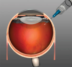
    Intravitreal injection of anti VEGFtreatment involves having a drug injected into your eye to treat certain retinal conditions that cause abnormal blood vessels to grow and leak under the retina. Patients with these conditions can lose central vision when abnormal blood vessels bleed or leak fluid under the retina at the back of the eye. A series of injections of anti-VEGF medicines are given into the back of your eye to stop these blood vessels from growing and help control the leakage. This treatment is highly effective in preserving central vision in many people.

    Which conditions require treatment with anti VEGF injections?

    • Wet age-related macular degeneration
    • Myopic choroidal neovascularization
    • Diabetic macular oedema
    • Retinal vein occlusion
    • Any other retinal conditions that causesfluid to leak under the retina

    Age-related macular degeneration (AMD)

    Age-related macular degeneration (AMD) is the leading cause of vision loss in people aged 50 years or older. It involves damage to the part of the eye called the macula. The macula is a small, but extremely important area located at the centre of the retina, the light-sensing tissue that lines the back of the eye.

    The macula is responsible for seeing fine details clearly. A person with AMD loses the ability to see fine details, both close-up and at a distance. This affects only the central vision. The side, or peripheral, vision usually remains normal. For example, when people with AMD look at a clock, they can see the clock’s outline but cannot tell what time it is.

    What is different about “wet” AMD?

    There are two types of AMD. Most people (about 75%) have a form called “early” or “dry” AMD, which develops when there is a waste build-up under the macula. This state is compatible with normal or partially reduced vision. A minority of patients with early AMD can progress to the vision-threatening forms of AMD called lateAMD. The most common form of late AMD is “exudative” or “wet” AMD. Wet AMD occurs when abnormal blood vessels grow underneath the retina. These unhealthy vessels leak blood and fluid, which can prevent the retina from working properly. Severe damage leads to severe permanent loss of central vision, but the eye is not usually at risk of losing all vision (going ‘blind’) as the ability to see in the periphery remains. There is a less common form of late AMD called geographic atrophy, where vision is lost through the macular tissue becoming completely worn out and there are no leaking blood vessels. Unfortunately, anti-angiogenic medicines cannot help this form of late AMD.

    Myopic choroidal neovascularization:

    This condition occurs in people who are highly myopic (short-sighted). When someone is highly short-sighted, the retina at the back of the eye is stretched due to the increased size of the eye. This stretching can make the retina thinner and prone to splitting. When this occurs, blood vessels from the choroid (the layer of the eye behind the retina) can grow underneath the retina. These new vessels (neovascularisation) leak blood and fluid, which can prevent the retina from working properly. Severe damage leads to severe permanent loss of central vision.


    What treatment is available?Diabetic macular oedema (DMO)

    Diabetic macular oedema is an eye condition occurring in people with both type 1 and type 2 diabetes. Macular oedema is swelling and thickening of the macula. The macula is a small area in the centre of the retina that contains a rich collection of nerve cells sensitive to light, fine detail, and colour. DMO occurs as a result of changes in retinal blood vessels in people with diabetes. Diabetes is characterised by increased levels of sugar (glucose) in the blood stream. Consistently high blood sugar can damage blood vessels, with the first signs appearing in the smallest vessels, called capillaries. The damaged blood vessels will leak, causing the build-up of excess fluid (oedema) and blood in the macula. It can lead to severe impairment of central vision in the affected eye.

    Retinal Vein Occlusion (RVO)

    RVO occurs when one of the retinal veins is blocked. The retina is the light sensitive tissue that lines the back of the eye and is responsible for the eyesight. RVO usually occurs when a retinal vein is: “pinched off” through the pressure of an artery lying on top of the vein; or is clogged with a blood clot or atherosclerotic plaque (fatty deposit in the wall of the artery); or is blocked by some inflammatory conditions.


    To Know more if you are a candidate for treatment kindly consult our doctor.